Neural Probes: What They Are, How They Work, and How to Choose One
A neural probe is an implantable device that carries recording or stimulating sites into neural tissue: the four types, how one records, and how to choose.
18 min read
Updated November 5, 2026

NeuroNexus · neuronexus.com · Resources
A neural probe is an implantable device that carries one or more recording or stimulating sites into neural tissue, so that the activity of neurons can be measured, or driven, at a known position. Most are electrical; newer designs add optical and chemical channels. This guide sets out how a probe records, the four types, acute and chronic use, optogenetics, how a probe is chosen, and the history of the device, with the published record behind each.
Research use only. NeuroNexus products and the procedures described here are for non-clinical research use, and must not be used in human or veterinary medical procedures.
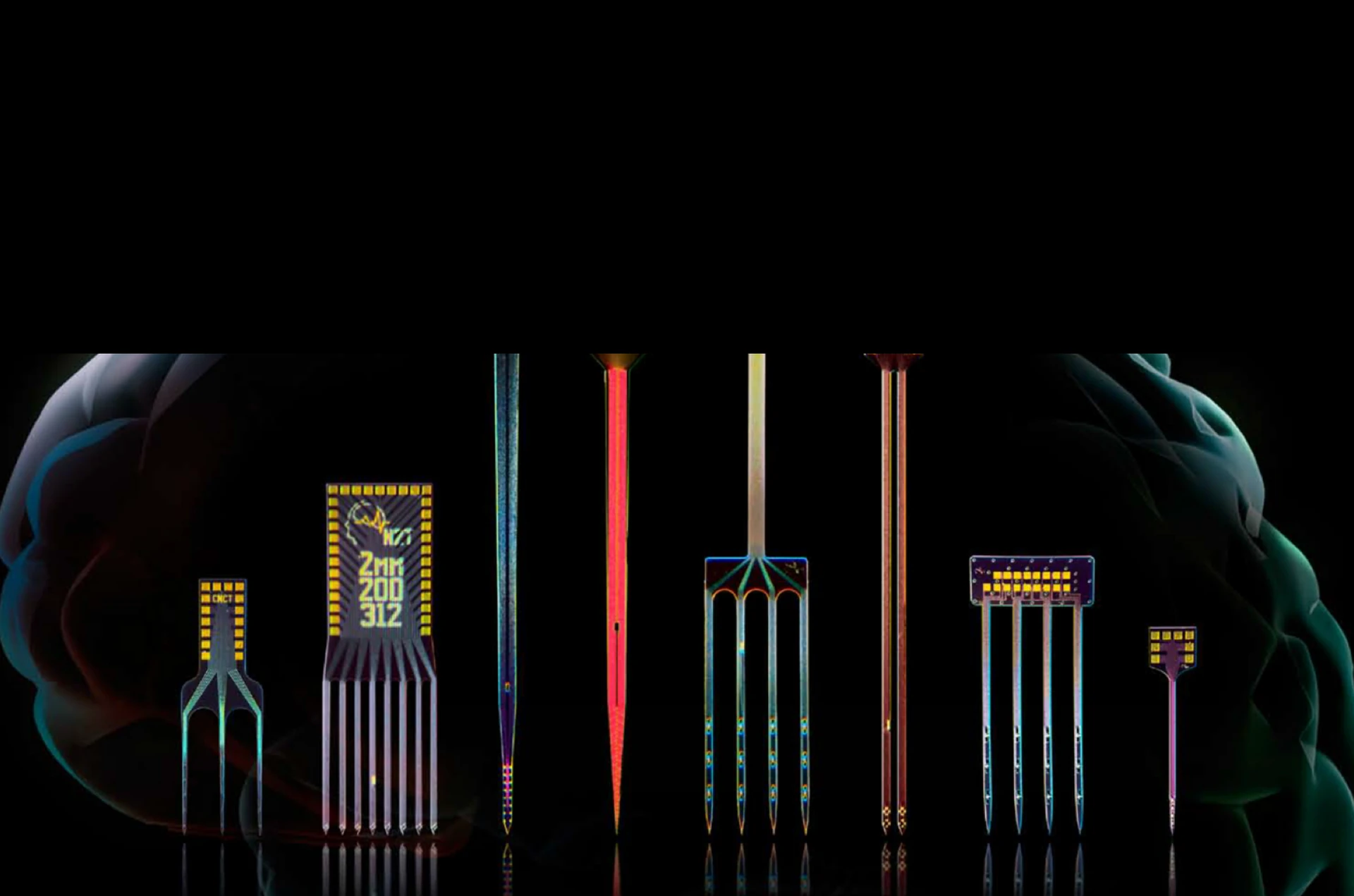
Figure 1. Silicon neural probes from one shank to eight. Each site sits at a position fixed by the design, so the recording is made where the design says it is.
What is a neural probe?#
A neural probe is an implantable device that carries one or more recording or stimulating sites into neural tissue, so that the activity of neurons can be measured, or driven, at a known position. The site is the working part: a small conductor, typically a few to a few hundred square micrometers, that forms an electrochemical interface with the extracellular fluid. The shank, wire, or film that carries the site into the tissue is the rest of the probe, and it ends in a connector that a headstage amplifies from. A probe with one site is a microelectrode; a probe with many sites at defined positions is a microelectrode array, and in current use "neural probe" means the array.
Most neural probes are electrical. They record the extracellular potentials that neurons produce, and on sites rated for it they deliver current for microstimulation. Newer designs carry other channels on the same shank: an optical fiber or a light source for optogenetics, a microfluidic channel for drug delivery, or a sensor for a chemical species. The probe is one of three parts of a recording chain. The other two are the headstage that amplifies and digitizes the signal and the data acquisition system that stores it; a probe package joins the probe to the headstage.
How does a neural probe record?#
A neural probe records the extracellular voltage at each site relative to a reference, and the geometry of the sites decides which neurons are heard and how well. A neuron firing an action potential drives a current through the extracellular space, and that current produces a small voltage at a site within about 100 micrometers of the cell body: the spike, tens to a few hundred microvolts, lasting about a millisecond. The slower sum of currents from many neurons, the local field potential, appears on every site in the region at up to a few millivolts. Both arrive at the same site; the amplifier's filter separates them, spikes above about 300 Hz and the field potential below.
Three properties of the site set the quality of what is recorded. Site area sets the trade between selectivity and noise: a smaller site hears fewer neurons and isolates them better, and has a higher impedance. Impedance, the site's opposition to current at the frequencies of a spike, sets the thermal noise the interface adds and how much of the signal reaches the amplifier; a typical 177 µm² iridium site measures 200 kΩ to 1 MΩ at 1 kHz. A coating that raises the electrochemical area without changing the footprint lowers the impedance at the same selectivity: NeuroNexus Z-Coat lowers site impedance by roughly an order of magnitude and is mechanically stable through repeated insertion, so the noise floor it buys holds across sessions. Site layout decides what the array as a whole can resolve: sites in a line along the shank profile a structure by depth; sites clustered 20 to 25 µm apart record the same neuron on several of them, which is what a spike sorter uses to separate units (Buzsáki (2004) ↗).
The reference closes the circuit. A site's voltage is measured against a reference electrode, a screw in the skull, a wire in the tissue, or a large site on the probe itself, and the choice of reference decides what is common to every channel and therefore what is subtracted. The shank carries the sites and the traces that connect them, and its width and thickness set how much tissue is displaced on the way in.
What are the types of neural probe?#
There are four types of neural probe in current use: micromachined silicon probes, wires and tetrodes, flexible polymer probes, and CMOS active-pixel probes, and they differ in how the sites are made and how the signal leaves the shank.
Silicon probes. A silicon probe is made by thin-film microfabrication: conductive and dielectric (insulating) layers deposited on a silicon wafer and patterned by photolithography, so that every site sits at a position fixed by the mask to sub-micrometer precision, the same on every probe of a design. The lineage is the Michigan probe, developed at the University of Michigan from 1970. NeuroNexus spun out of that program in 2004 and offers the largest catalog of the type: more than 210 designs with 4 to 1,024 sites on 1 to 16 shanks, 1.5 to 60 mm long, on 15 or 50 µm silicon, with iridium, platinum, SIROF, and Z-Coat site options, and custom geometries against a target the catalog does not cover. The site metal and the dielectric are chosen for the recording and for the years the probe spends in tissue. The catalog is at silicon neural probes.
Wires and tetrodes. A microwire is a single insulated metal wire with an exposed tip; a tetrode is four wires twisted together so that one neuron appears on all four at slightly different amplitudes. Wires are inexpensive and robust, and a tetrode isolates units well at one depth. What they lack is defined geometry: the position of each tip is set by hand at assembly rather than by a mask, so the layout varies from array to array and cannot sample a structure in a known pattern. The silicon equivalent of the tetrode is a site cluster at a fixed pitch, and NeuroNexus makes those as the polytrode and tetrode layouts.
Flexible polymer probes. A polymer probe places the sites on a thin polyimide or parylene film rather than on silicon, so the array bends with the tissue. On a surface the film is a thin-film grid, the surface counterpart of the silicon probe: NeuroNexus makes 53 designs for the cortical surface, the skull, the heart, and peripheral nerves, from 3 to 256 sites. In tissue a polymer shank is less stiff than the tissue's own movement, which lowers the chronic response at the cost of needing a stiffener or a carrier to insert.
CMOS active-pixel probes. An active-pixel probe puts an amplifier and a digitizer under every site, so the shank carries a digital data stream rather than one wire per site, and a single shank holds hundreds to a thousand sites. Neuropixels, made by imec, records 384 channels chosen from 960 to 5,120 sites, with the circuit in the probe base (Jun et al. (2017) ↗). SiNAPS, commercialized by NeuroNexus as the HD Series, puts the pixel under the site itself and records every one of its 256 or 1,024 sites at once, at 12 bits and 20 kS/s per pixel (Angotzi et al. (2019) ↗). Active-pixel probes record only; stimulation goes through a passive probe.
How are neural probes used acutely and chronically?#
An acute recording lasts hours on an anesthetized or head-fixed animal; a chronic recording lasts weeks to months on an implanted animal, and the two demand different things from the probe and its package. In an acute session the probe is held by a micromanipulator, lowered to the target, and withdrawn at the end, so the design can be long, stiff, and carried on a rigid package with a standard connector; the same probe is cleaned and used again. In a chronic preparation the probe stays in the tissue, and what matters is the package the animal wears and what the tissue does around the shank.
The package decides the chronic preparation. A chronic build puts the connector on a flexible cable, so the array floats with the brain rather than being held rigid against the skull, or on a compact assembly light enough for a mouse. A microdrive adds the ability to move the probe after implantation: tissue settles over the first weeks and units drift out of the sites' reach, and a drive advances the probe a few tens of micrometers so the recording resumes. NeuroNexus makes six microdrives; the dDrive carries the chronic silicon-probe packages, and the oDrive adds an optical fiber for chronic optogenetics. The choice of package and drive is described in Choosing a Probe Package.
The tissue responds to any implant. Glial cells encapsulate the shank over the first weeks and the neurons nearest it can withdraw, which raises impedance and lowers the amplitude of units. The response scales with the mass and stiffness of what is implanted and with how the shank moves relative to the tissue, which is why a thin shank, a floating package, and a small, light headstage matter more in a chronic preparation than any other specification. NeuroNexus reported the first chronic performance of silicon probes in cortex (Kipke et al. (2003) ↗; Vetter et al. (2004) ↗) and showed that a thin lateral platform reduces the encapsulation around the sites (Seymour & Kipke (2007) ↗); the 15 µm substrate in the catalog comes from that work.
Can a neural probe be used for optogenetics?#
Yes: an optoelectrode carries an optical fiber alongside the electrode array, so light is delivered on the same trajectory as the recording sites, and the neurons that are recorded are the neurons that are stimulated. NeuroNexus builds the optrode package on most of its package series: the fiber is fixed at assembly, terminates in a standard fiber-optic connector for a separate laser or LED, and is confirmed on the quotation by its core, cladding, numerical aperture, and length. The light pulse is timestamped with the neural data through the acquisition system's digital input, so pulse onset and offset are known to the sample.
Light on a metal site produces a voltage transient of its own, the photoelectric artifact: photons release charge at the metal surface, the illuminated interface shifts its potential, and the absorbed energy heats it, and the amplifier records the sum, time-locked to the pulse, in the same milliseconds as the response (Cardin et al. (2010) ↗; Kozai & Vazquez (2015) ↗). Its size is set by the irradiance at the site, the exposed metal area, the site interface, and the pulse edges. NeuroNexus removes it at the electrode, at two levels: Z-Coat changes the site interface so the transient is suppressed, and the optrode package places the fiber so the sites are not lit directly. The mechanism and the remedies are set out in Photoelectric Artifact in Silicon Probes.
How is a neural probe chosen?#
A neural probe is chosen by fitting its geometry and its package to the target, the measurement, the duration of the experiment, the species, and the recording system, in that order; function follows fit. Six decisions make the choice.
Species and depth set the shank length and, with it, the stiffness the insertion needs: a mouse cortex is reached at 1 to 2 mm, a rat hippocampus at 3 to 4 mm, a primate structure at 20 mm or more, and the catalog's 1.5 to 60 mm lengths cover them on a 50 µm substrate where stiffness is needed and 15 µm where the tissue response matters more. The measurement decides the site layout: a laminar profile wants sites in a line at the pitch of the layers; unit isolation wants clusters of closely spaced sites; a map across a structure wants several shanks at a known separation; a study of the tissue on both sides of a shank wants a double-sided design, which NeuroNexus makes as the Fusion probe, with sites on both faces. Channel count follows from the layout and the structure, from 4 sites on a single shank to 1,024 on an active-pixel probe, and the count fixes the headstage and the connector. Acute or chronic sets the package: a rigid package and a micromanipulator for a session, a floating or compact package and a microdrive for an implant. Optogenetics adds the fiber as a package option and makes Z-Coat the site choice. Insertion closes the loop: the shank's cross-section and the tissue's own movement set what the design tolerates, and a thin shank is inserted with a carrier where it would otherwise buckle.
| Type | Sites per probe | Geometry | Chronic use | Stimulation | Optogenetics | Typical subjects |
|---|---|---|---|---|---|---|
| Silicon (micromachined) | 4 to 256 passive | fixed by the mask; linear, cluster, multi-shank, double-sided | acute and chronic; floating packages and microdrives | yes, on rated sites | optrode packages | mouse to non-human primate |
| Wire and tetrode | 1 to a few dozen | set at assembly | chronic, with a drive | yes | separate fiber | rodent |
| Flexible polymer | 3 to 256 on a film | fixed by the mask; conformal | chronic surface recording; penetrating with a carrier | yes | separate fiber | mouse to large animal |
| CMOS active-pixel | 256 to 1,024 recorded at once (SiNAPS); 384 chosen from 960 to 5,120 (Neuropixels) | fixed by the mask; dense linear | acute and chronic | no | separate fiber; optrode build on SiNAPS | rodent; non-human primate versions |
The Electrode Finder applies these decisions to the catalog: it takes the site count, pitch, depth, and shank layout the experiment needs and shows which designs fall inside those ranges. Every design has a datasheet with the packages it is built in and the headstage each package mates, and the packages themselves are set out in Choosing a Probe Package.
A short history of the neural probe#
The neural probe as an array with sites at defined positions dates from 1970, when the first microelectrode made by integrated-circuit methods was reported at Stanford and then developed at the University of Michigan into the planar multisite silicon probe. Wise, Angell, and Starr patterned a recording site on a silicon substrate with the lithography of the integrated circuit, and showed that the methods of the chip could make an electrode (Wise et al. (1970) ↗). Najafi, Wise, and Mochizuki reported a multichannel recording array made with a high-yield, circuit-compatible process, the form every silicon probe since has taken (Najafi et al. (1985) ↗). The device was known as the Michigan probe; NeuroNexus was founded in 2004 by members of that program, Daryl Kipke, Rio Vetter, and Jamille Hetke, to make it available to the field, and reported the first chronic recordings of neuronal spike activity from silicon probes in cerebral cortex (Kipke et al. (2003) ↗; Vetter et al. (2004) ↗).
Buzsáki set out what large-scale recording of neuronal ensembles requires of the probe, many sites close enough together that one neuron appears on several, and the layouts that carry his name followed (Buzsáki (2004) ↗). The active-pixel probe arrived in the 2010s, with the amplifier and digitizer on the probe: Neuropixels put the circuit in the probe base and 960 sites on one shank (Jun et al. (2017) ↗), and SiNAPS put a pixel under every site so that all of them record at once (Angotzi et al. (2019) ↗). NeuroNexus silicon probes have been used in more than 5,000 peer-reviewed publications; the citation database lists them by design.
Questions and answers#
What is a neural probe?#
A neural probe is an implantable device that carries one or more recording or stimulating sites into neural tissue, so that the activity of neurons can be measured, or driven, at a known position. Most are electrical; newer designs add optical and chemical channels on the same shank.
What is the difference between a silicon probe and a wire electrode?#
A silicon probe has its sites at positions fixed by a lithographic mask, the same on every probe of a design, so an array can sample a structure in a known pattern; a wire electrode's tip is placed by hand at assembly, so its position varies from array to array. A tetrode, four wires twisted together, isolates units well at one depth; the silicon equivalent is a cluster of sites at a fixed 20 to 25 µm pitch.
How many channels does a neural probe have?#
From one site on a microwire to 1,024 on an active-pixel probe. NeuroNexus passive silicon probes have 4 to 256 sites on 1 to 16 shanks; SiNAPS active-pixel probes record 256 or 1,024 sites at once; Neuropixels probes record 384 channels chosen from 960 to 5,120 sites. The count follows from the site layout the measurement needs, and it fixes the headstage and the connector.
How long does a chronic neural probe last?#
A chronic implant records for weeks to months. Over the first weeks the tissue settles and glial cells encapsulate the shank, so impedance rises and unit amplitude falls; a thin shank, a floating package, and a light headstage reduce the response, and a microdrive recovers units by advancing the probe a few tens of micrometers. NeuroNexus reported the first chronic performance of silicon probes in cortex in 2003 and 2004.
Can a neural probe be used for optogenetics?#
Yes. An optrode package fixes an optical fiber alongside the array, so light is delivered on the trajectory of the recording sites. Light on a metal site produces the photoelectric artifact; NeuroNexus Z-Coat suppresses it at the site, and the optrode package places the fiber so the sites are not lit directly.
How do I choose a neural probe?#
By fitting the probe to the target, the measurement, the duration, the species, and the recording system, in that order: species and depth set the shank length, the measurement sets the site layout, the layout sets the channel count, acute or chronic sets the package, and optogenetics adds the fiber. The Electrode Finder applies those decisions to the catalog.
Related documents#
- Silicon neural probes, the catalog
- NeuroNexus Silicon Probes and Thin-Film Grids: An Introduction
- Choosing an Electrode for Your Experiment
- Choosing a Probe Package
- NeuroNexus Probe Technology: Questions and Answers
- Photoelectric Artifact in Silicon Probes
- Neuropixels, SiNAPS and Silicon Probes: How to Choose
Citations#
- Wise, K. D., Angell, J. B., and Starr, A. (1970). An integrated-circuit approach to extracellular microelectrodes. IEEE Transactions on Biomedical Engineering 17(3), 238–247. 10.1109/TBME.1970.4502738 ↗
- Najafi, K., Wise, K. D., and Mochizuki, T. (1985). A high-yield IC-compatible multichannel recording array. IEEE Transactions on Electron Devices 32(7), 1206–1211. 10.1109/T-ED.1985.22102 ↗
- Kipke, D. R., Vetter, R. J., Williams, J. C., and Hetke, J. F. (2003). Silicon-substrate intracortical microelectrode arrays for long-term recording of neuronal spike activity in cerebral cortex. IEEE Transactions on Neural Systems and Rehabilitation Engineering 11(2), 151–155. 10.1109/TNSRE.2003.814443 ↗
- Vetter, R. J., Williams, J. C., Hetke, J. F., Nunamaker, E. A., and Kipke, D. R. (2004). Chronic neural recording using silicon-substrate microelectrode arrays implanted in cerebral cortex. IEEE Transactions on Biomedical Engineering 51(6), 896–904. 10.1109/TBME.2004.826680 ↗
- Buzsáki, G. (2004). Large-scale recording of neuronal ensembles. Nature Neuroscience 7(5), 446–451. 10.1038/nn1233 ↗
- Seymour, J. P., and Kipke, D. R. (2007). Neural probe design for reduced tissue encapsulation in CNS. Biomaterials 28(25), 3594–3607. 10.1016/j.biomaterials.2007.03.024 ↗
- Cardin, J. A., Carlén, M., Meletis, K., Knoblich, U., Zhang, F., Deisseroth, K., Tsai, L.-H., and Moore, C. I. (2010). Targeted optogenetic stimulation and recording of neurons in vivo using cell-type-specific expression of Channelrhodopsin-2. Nature Protocols 5, 247–254. 10.1038/nprot.2009.228 ↗
- Kozai, T. D. Y., and Vazquez, A. L. (2015). Photoelectric artefact from optogenetics and imaging on microelectrodes and bioelectronics: new challenges and opportunities. Journal of Materials Chemistry B 3, 4965–4978. 10.1039/C5TB00108K ↗
- Jun, J. J., et al. (2017). Fully integrated silicon probes for high-density recording of neural activity. Nature 551, 232–236. 10.1038/nature24636 ↗
- Angotzi, G. N., et al. (2019). SiNAPS: An implantable active pixel sensor CMOS-probe for simultaneous large-scale neural recordings. Biosensors and Bioelectronics 126, 355–364. 10.1016/j.bios.2018.10.032 ↗
Further examples of neural probes in published research are in the NeuroNexus citation database.